Embryonic stem cells (ESCs) are isolated from the inner cell mass of an embryo and can differentiate into all types of cells making up an organism. Thus, the study of ESCs is important to understand the basic science of developmental biology and for the practical application of creating cell lines, tissues and organs that can be used in the fields of tissue engineering, and drug discovery. As a stem cell differentiates down various lineages into various cell types its communication tasks become more complex and so must its glycome to facilitate cell-cell and cellextracellular matrix interactions. Furthermore, the glycans in extracellular matrix form a glyconiche that can serve to control the growth and differentiation of a stem cell. Work in the BCME Constellation at Rensselaer is focused on understanding how the glycome changes as ESCs differentiate and how the glyconiche of a stem cells control their growth and differentiation. Changes in glycosaminoglycans (GAG) structure, which are long and linear polysaccharides consisting of repeating disaccharide unit, are of interest. They found attached to protein core, comprising proteoglycans (PG) and found on cell surface and extracellular matrix.
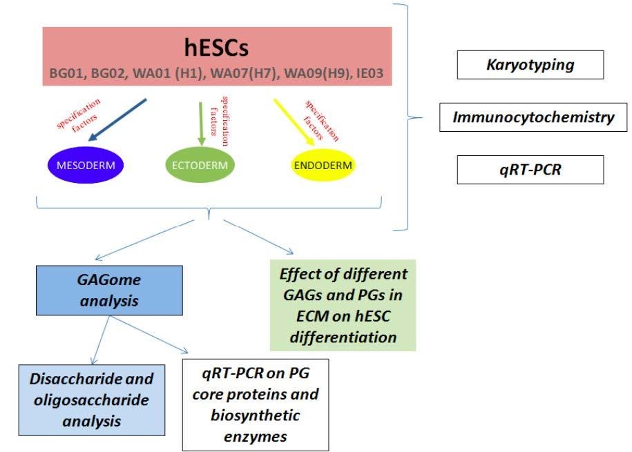
Schematic plan of glycan analyses upon differentiation of several human Embryonic Stem Cells lines into representatives of mesoderm, endoderm and ectoderm.
Comprehensive study of glycomics of ESC is accomplished on different levels including transcriptomic, proteomic and structural analyses (Figure 2). Pluryipotent ESCs are driven to differentiate into representatives of different germ layers and GAGome changes are monitored along the differentiation pathways. Meanwhile, qRT-PCR is used to detect expression level of GAG biosynthetic enzymes (Figure 3), which are later confirmed by western blotting. LC-MS and CE electrophoresis are used to determine the structure of GAGs.
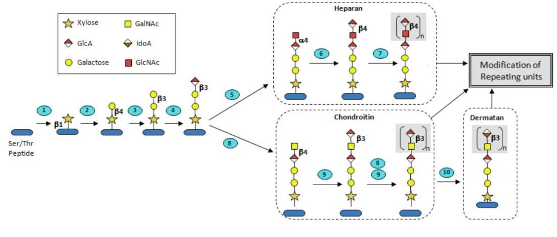
Biosynthetic pathway of heparin/heparan sulfate and chondroitin sulfate/dermatan sulfate glycosaminoglycans. The Ser/Thr-containing polypeptide core protein is glycosylated and modified through the series of enzymatic reactions occurring in the endoplasmic reticulum and Golgi apparatus. Numbers on steps correspond to enzymes catalyzing relevant reactions as (1): XYLT1, XYLT2, (2): B4GALT7, (3): B3GALT6, (4): B3GALT3, (5): EXTL2, EXTL3, (6): EXT1, EXT2, (7): EXT1, EXT2, EXTL1, EXTL3, (8): CSGALNACT1, CSGALNACT2, (9): CHSY1, CHPF1, CHSY3, CHPF2, and (10): DSE1, DSE2 (Nairn, A. V., Linhardt, R. J. et al, 2007, J. Proteom. Res.)
The effect of glycan structure on stem cell fate is determined using cell-based microarray technologies, which allows high throughput analyses of ESCs. Cell-based microarray can be coupled with in-cell immunofluorescence to determine the effect of various signaling molecules on stem cell state
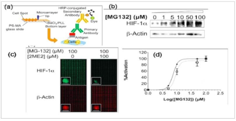
Three-dimensional cellular microarray for the study of murine stem cells. a. approach for microarray in-spot immunofluorescence for protein expression; b. Western blot for comparison of up-regulated expression to expression; c. determination of HIF-1α expression expression;quantification (Biotechnol Bioeng. 2010, 106:106-118.) HIF-1 α actin in spot to actin and d. of data.